


The disease is less studied, although several thousand observations with subsequent diagnosis and treatment have been described.
Many diversity and non -specific clinical pictures of pelvic varicose veins lead to a rough error from the diagnosis, which in the future affects the consequences.
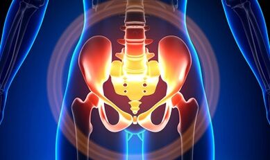
Characterization of small pelvic varicose veins
Pelvic veins are several times longer than the artery, which determines their large capacity.This is due to the phylogenesis of the vascular system of the pelvic region.Pelvic veins have high adjustments and potential to restructure, which contribute to the formation of a compact network.
The speed and direction of blood flow are controlled by the valve, which is controlled by complex humoral mechanisms.The valve offset the pressure on different parts of the venous network.
When the valve stops performing their function, blood stagnation develops, leading to blood vessel pathology and the formation of varicose veins.The specialty of small pelvic veins lies in the fact that the uterine ligaments, which support the lumen of the vessel, can also narrow it, causing pathology.
Cause
The pathology of the pelvic vein pathology may be due to the following reasons:
- Infringement of blood exit;
- Barrel Venous Prison;
- Compression of collateral stems by changed uterus position, for example, in retroflexia;
- Valve ovarian failure (congenital or acquired);
- Post Syndrome -Flebitical Obstructive;
- Connective tissue pathology;
- Arteriovenous angiodisplasia;
- Prolonged sitting, hard physical labor;
- Varicose veins from the lower leg;
- Pregnancy (3 or more) and giving birth (2 or more);
- Diseases of female genitalia (chronic salpingoophorites, ovarian tumors, uterine fibroids and genital endometriosis);
- Pelvic organs adhesive process;
- Obesity.
Classification by degree of disease
In terms of expanded veins, the following degrees are distinguished:
- up to 0.5 cm, vascular stroke "corkscrew";
- 0.6-1 cm;
- More than 1 cm.
The choice for the disease
- perineum varicose veins and vaginal vestibules;
- venous syndrome full of pelvis;
Symptom
- The most common pain - frequent in the lower abdomen, perineum after long and dynamic static overstresspresses.Pain is increasing in the second phase of the cycle, after hypothermia, fatigue, stress, hunting of various diseases.
- The feeling is "uncomfortable", it hurts with sex and after that.
- Dysmenorrhea - menstrual disorders, including pain syndrome.
- Secrets, more than normal, galls of genital tract.
- Blood gestures lead to infertility, inability, termination of pregnancy.
- Urinary tract infringement due to the development of the bladder vein.
Diagnostics
Diagnosis of the disease only by successful complaints in only 10 % of cases.
Palpation of the inner wall of the pelvis, allowing to feel the oval seal and the vein node.When examined in the mirror, the cyanosis of the vaginal mucous membrane can be seen.
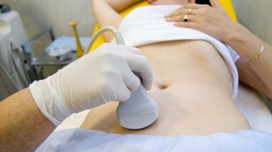
The procedure for choosing is an ultrasound study with color doppler mapping, which allows you to identify not only the extension of ovarian veins, but also venous thrombosis, posttrombopbititis occlusion.With ultrasound, it can be seen, "worm", a structure without reflection of signals, locally on the surface of the uterus.
The effects of the Doppler study are based on "colors" in blue and red, venous blood and arteries, which match.
Tools for ultrasonic examination using special programs recognize blood movement from sensors and in other directions, calculating blood flow velocity and vessels.
But the actual definition of veins or arteries remains behind the doctor.The doppler method works in almost all cases, our body determines the exception to the rules, because the blood flowing from the liver is not always the artery and vice versa.
Therefore, ultrasonic diagnostic doctors see an artery or vein, its size, blood flow rate and many indications that ordinary people do not need, but play an important role in making diagnosis.To do this, use transabdominal and transvaginal sensors.
In 5.7% of cases, the disease was accidentally recognized during examination.Typically, the diameter of the ovaric vein is 0.4 cm.
CT and MRI have great accuracy.Using this method, you can find the accumulation of varicose veins in the ligaments of the uterus, ovaries and around these organs.It is possible to determine the same pathology.
A very reliable method is a phloographic study.
Instead, it is performed at the height of the Valsalva sample, against blood flow.This allows you to see the valve failure.
Retronenoscopy on the left, kidney phlebography, super -sequenic and phlebovariography phlebovarioscopy in both sides are also used.This method allows you to determine the changes in hemodnamese and anatomy in the kidney veins and places that fall into the gonadny veins.
Super -spent phlebovarioscopy is performed by catheterization of gonad veins via counterattotal or subclavian veins, with the introduction of subsequent contrast.
Most blood from the varicose hornless plexus is removed through the ovarian veins.But in hypertension, it occurs through the uterine veins that are not adjusted into the inner iliac vein.Plexus veins, where outflows can occur, including sacral and bladder plexus.
In Phlebo -icigography, 3 stagnation stagnation stages are distinguished in left ovarian cluster plexus:
- There is no outflow from the left ovary plexus or it is done along the additional short road.
- There is even further.
- Two additional outflow pathways or one additional and additional can be seen.
At levels 2 and 3, the right ovarian cluster varicose veins are formed.
Laparoscopy is used for differential diagnosis.The complicated pathological veins are in the ovaries, towards round and wide ligaments.They look like large cyanotic conglomerate with thin and tense walls.
The complexity of the diagnosis lies in the fact that the disease is often hidden behind the signs of inflammation, distinguished by clinical manifestations, masked under endometriosis, internal organ prolapse, postoperative neuropathy and many extragenital diseases.
Treatment
The main goal of treatment is to eliminate reflux in the veins.In the early stages of the disease, conservative treatment was used.In the final stages of the disease, the treatment of choice is surgery.
Conservative treatment

It consists of normalizing veins, hemodynamic increases and trophic processes.
Symptomatic treatment to eliminate individual symptoms.Nonsteroidal anti -inflammation for pain, with bleeding - hemostatic therapy.
The main medicines in conservative treatment are venotonic and antiplatelet.
Phlebotonics - improves vascular wall tone and improves blood flow.With this disease, it is best to consult a gynecologist about certain medications.
An important method is physiotherapy training.
Surgical
- Varicose veins of resurgence.
- Gonado-Cavalry Shunting.
- Sclerosis for laparoscopy.
- Occrames ovarian veins use x-ray-endovascular methods.
The recovery of the people
Because the main thing in the occurrence of the disease is the weakness of the valve, all folk remedies used for the development of varicose veins from the bottom of the foot are also used for this pathology.
The most commonly used: regular hazel, hop, nettles, horse chestnut, dandelion root, tea mushrooms, willow, oak, wort St.John, series, flower pollen and many more growing.
Effective are: Treatment with oak baths, chestnuts, willow, chamomile, pharmacy, drying herbs, St.John.
Prevention
- The first thing to do if there are complaints, predictors or illnesses listed above - contact a gynecologist.
- It is necessary to normalize the work regime and rest, try not to stay in a position for a long time, physical tension.
- Exercise for the prevention of "pedal", "stand-up stack", "feet"
- Comply with diet: eat foods with vitamins E, R, C, try to eat only white meat, less fat, replace them with fruits, vegetables, cereals.
- Drink sufficient amount of fluid, but not less than 1.5 liters per day.
- Losing excess weight, bad habits.
- Consulting with the attending physician wearing compression linen, it will increase blood flow from the lower leg, thus less stagnation in the pelvis.
- Avoid shower, sauna, steam room, hot bath.
In order to avoid pain with difficult illnesses, it is necessary to follow the preventive suggestions listed above.Treat your health as the most valuable in life.
On a little suspicious symptoms that you can't get rid of for a few days, you should consult your doctor.He must give you qualified help and save you from suffering.